Synthesis and characterization of copper peroxide
The as-prepared copper peroxide (CP) nanospindles have been synthesized in accordance with an method reported beforehand with minor modifications. The outcomes exhibited that CP nanoparticles offered a uniform spindle-like morphology (Fig. 1A) with a DLS measurement of 131.30 ± 1.87 nm (Fig. 1B). The polymer dispersity index (PDI) of CP nanospindles was round 0.10, suggesting good dispersibility in aqueous answer. The X-Ray diffraction spectrum verified the crystal morphology of CP nanospindles (Fig. 1C). The X-ray photoelectron spectrum revealed the presence of Cu and O parts in CP nanospindles (Fig. 1D). The spectrum of Cu2p featured with two typical peaks at 928.58 eV and 952.58 eV from Cu2+ 2p1/2 and Cu2+ 2p3/2 accompanying with two satellite tv for pc peaks at 961.95 eV and 941.5 eV (Fig. 1E). In the meantime, two peaks in O 1s spectrum at 530.7 and 532.5 eV have been deconvoluted to C = O plus O-O, respectively, hinting on the existence of PVP plus peroxo teams (Fig. 1F). A permanganate (MnO4−) primarily based colorimetric technique was utilized to analyze the presence of peroxide bonds in CP nanospindles. Typically, magenta MnO4− may very well be lowered to colorless Mn2+ ions by H2O2. Just like H2O2, the colour of MnO4− answer pale away quickly by the CP nanospindles whereas CuO confirmed no such phenomenon, indicating the presence of peroxide bonds in CP nanospindles (Fig. 1G).
Inefficient intracellular H2O2 content material is without doubt one of the main boundaries to realize passable chemodynamic remedy (CDT) efficacy. To surmount these obstacles, researchers have give you varied optimized CDT brokers. Catalysts composed of self-supplied H2O2 and Fenton response exercise have attracted a lot consideration. A colorimetric technique primarily based on the precept that Ti4+ and H2O2 reacted to type yellow peroxide-titanium complicated was employed to analyze the H2O2 self-supplying efficiency of CP nanospindles. Within the presence of H2O2, a yellow precipitation composed of peroxide-titanium was produced upon the response of H2O2 and titanium ions. As proven in Fig. 1H, H2O2 may very well be effectively produced at pH 5.5 whereas no H2O2 was detected when pH worth elevated to 7.4, implying the pH responsive H2O2 technology efficiency of CP nanospindles. As well as, with the focus of CP nanospindles growing to 100 µg/mL, the focus of H2O2 produced elevated to ~ 235.56 µM (Fig. 1I and S1a), suggesting that CP nanospindles might function an environment friendly exogenous H2O2 repository. In distinction, CuO nanoparticles couldn’t induce H2O2 manufacturing (Fig. 1I and S1b).
5,5’-Dithiobis-(2-nitrobenzoic acid) (DTNB), an Ellaman agent, is used to analyze the GSH depletion efficiency of CP nanospindles. The attribute peak at 412 nm of DTNB progressively disappeared, implying the wonderful GSH depletion efficiency of CP nanospindles was effectively (Fig. 1J). Furthermore, Cu2+ ions, lowered to Cu+ ions by GSH, might function a extra environment friendly Fenton catalyst in comparison with iron-based ones. Methylene blue was used as colorimetric probe to quantify the hydroxyl radicals (∙OH) manufacturing. With the GSH focus elevated from 0 to 4 mM, extra ∙OH was produced. Nevertheless, when the GSH focus elevated over 8 mM, the produced ∙OH was balanced by GSH, additional confirming the GSH depletion was necessary for ROS-based most cancers remedy (Fig. 1Ok).
Inspired by the GSH depletion and ∙OH manufacturing efficiency, we then explored the in vitro anticancer efficiency of CP nanospindles. As proven in Fig. 1L, greater than 80% most cancers cell loss of life was induced on the focus of 45 µg/mL of CP nanospindles, whereas solely ~ 60% cell viability lower induced by CuO nanoparticles, implying higher chemodynamic remedy efficacy as a result of introduction of self-supplying H2O2. Hereafter, the dose near the half maximal inhibitory focus of CP nanospindles, 15 µg/mL, was chosen for the next in vitro experiment. As well as, after cultured with CP nanospindles and CuO for six h, CT-26 cells have been stained with DCFH-DA, a ROS fluorescent probe. CT-26 cells incubated with CP exhibited considerably extra vivid inexperienced fluorescence in comparison with these with CuO nanoparticles, indicating extra hydroxyl radicals induced by CP nanospindles (Fig. 1M). Collectively, the as-synthesized CP nanospindles, exhibiting a uniform spindle-like morphology, signify an environment friendly CDT agent for tumor inhibition through catalyzed GSH consumption and ROS technology (Fig. 1N).

Synthesis, characterizations and in vitro anti-tumor efficiency of copper peroxide nanospindles. (A-C) TEM pictures (A), DLS measurement distributions (B) and XRD patterns of CP (C). (D–F) XPS spectrum evaluation of CP. (G) UV-Vis spectra evaluation for the existence of peroxo teams in CP nanoparticles. (H and I) UV-Vis spectra of Ti (SO4)2 and CP underneath varied pH values (H) and varied concentrations of CP (I). (J) GSH depletion by CP nanospindles after varied incubation time. (Ok) UV-Vis spectra demonstrating the degradation of Methylene blue (MB) by CP nanospindles with varied concentrations of GSH. (L) Cell viability evaluation of CT-26 cells uncovered to completely different concentrations (equal to copper ions) of CP or CuO for twenty-four h. The experiments have been repeated thrice, n = 3. (M) Fluorescence pictures of CT-26 uncovered to the completely different concentrations (15, 30 µg/mL) of CP or CuO for six h and subsequently stained by DCFH-DA (10 µM; 20 min). Cell nucleus have been stained by Hoechst 33,342 (HO; 10 µg/mL) for 20 min. (N) A schematic graph confirmed CP nanospindles might induce activatable ∙OH manufacturing and GSH consumption for enhanced chemical dynamic remedy
Autophagy induced by CP exhibited no tumor cell destiny dedication
Autophagy is a lysosome dependent and dynamic degradation pathway (Fig. 2A), that removes aberrant organelles, misfolded and long-live proteins, and invading pathogens, critically influencing pathogenesis and therapy of human illnesses [40, 41]. Basal-level autophagy is a important mechanism for sustaining cell stability and selling cell survival, occurring at low ranges in nearly all mammalian cells. A rise in autophagy degree is commonly noticed when cells uncovered to bodily, chemical, or organic stress [42]. Herein, the as-synthesized copper peroxide nanoparticles have been verified to induce autophagic flux. Autophagy associated protein LC3 was constitutively expressed and massively distributed in cytoplasm as LC3-I, nevertheless, upon autophagic induction, could be cleaved and conjugated to phosphatidyl ethanolamine to type lipidated LC3 (denoted as LC3-II), collaborating within the meeting of autophagosome (Fig. 2A). Subsequently, analyzing LC3 conversion from cytosol LC3 (LC3-I) to lipidated LC3 (LC3-II) by immunoblotting may very well be used to analyze autophagic induction. As displayed in Fig. 2B and C, the as-synthesized CP nanoparticles induced LC3 conversion, with concentration- and time- dependent patterns. To steer extra proof, CT26 cells over-expressing GFP tagged LC3 (denoted as GFP-LC3/CT26) have been constructed. The binding of GFP-LC3 to autophagosomal membranes, often noticed as inexperienced dots, is a dependable indicator for autophagosome formation [43, 44]. However, GFP-LC3 dots co-localization with lysosomes mirror autolysosome formation, important for the degradation of autophagic cargo [43, 44]. As noticed underneath a confocal microscope, CP might induce GFP dot formation, which exhibited an apparent co-localization with LysoTracker Crimson-indicated lysosomes (Fig. 2D). These outcomes demonstrated that CP nanoparticles might induce auto-lysosome formation with out disrupting autophagosome-lysosome fusion. Chloroquine (CQ), an antimalarial agent, additionally might block the downstream of autophagy course of by advantage of alkalizing lysosome and inhibiting autophagosome-lysosome fusion [45]. Herein, within the presence of CQ, CP might additional elevate LC3-II accumulation, indicating that CP actually induced autophagic induction reasonably than autophagic blockage (Fig. 2E). To sum up, the as-synthesized CP nanoparticles really elicited autophagy.
Autophagy induction is a trademark underneath varied pathological and pathophysiological situations, probably selling loss of life or survival, that are two distinct paradigms that decide cell destiny. Generally, autophagy provoked by nanomaterials promotes cell loss of life, however numerous nanomaterials can even set off survival-promoting autophagy [42, 44, 46,47,48]. Primarily based on the decisive function of nanomaterial-induced autophagy in tumor cell destiny, successfully avoiding or amplifying autophagy induced by nanomaterials could grow to be an growing new weapon towards cancers, particularly these which can be troublesome to deal with and drug-resistant [46, 48, 49]. Therefore, uncovering the dedication of CP-induced autophagy on tumor cell destiny is important for propelling CP-based tumor catalytic remedy. Nevertheless, our examine demonstrated that autophagy induced by CP offered no impact on cell destiny. Wortmannin was reported to dam autophagic initiation through inhibiting class III phosphatidylinositol 3-kinase (PI3K) and broadly used as autophagic inhibitors [50, 51]. Within the current examine, each western blot evaluation and the parallel statistical evaluation (Fig. 2F) indicated autophagy elicited by CP may very well be considerably attenuated by wortmannin. Nevertheless, autophagy inhibition by wortmannin wouldn’t have an effect on CP elicited cell loss of life (Fig. 2G), which indicated autophagy induced by CP exhibited no most cancers cell destiny choice. To offer extra proof, shRNA towards ATG5 was deployed to abrogate the expression of ATG5 in CT-26 most cancers cell line. ATG5 is a important element of autophagy, collaborating the regulation of autophagosome formation and enlargement [52]. Herein, immunoblotting and statistical outcomes demonstrated that the decreased expression of ATG5 might considerably suppress autophagic induction by CP (Fig. 2H). Nonetheless, the expression abrogation of ATG5 additionally confirmed no any important affect on CP elicited most cancers cell loss of life (Fig. 2I). Collectively, though the as-synthesized copper peroxide nanoparticles induced autophagy, the autophagy induced by CP had non affect on cell destiny dedication.
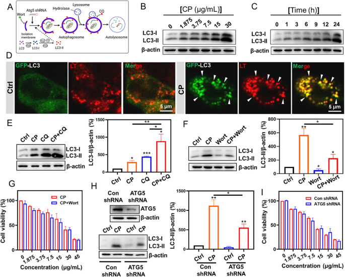
Autophagy induced by CP exhibited no tumor cell destiny dedication. (A) A schematic illustration of autophagy course of, throughout which, a number of autophagy inhibiting approaches have been additionally listed. Wort: Wortmannin; CQ: chloroquine. (B–C) Western blot evaluation of CT-26 after the incubation with completely different doses of CP for twenty-four h (B), or handled with CP (15 µg/mL) for the indicated occasions (C). (D) Confocal fluorescence pictures of LysoTracker™ Crimson (LT) stained GFP-LC3/CT-26 cells after the therapy with CP (15 µg/mL) for twenty-four h. Lysosomes have been indicated by LT (75 nM) staining for 15 min. (E) Western blot evaluation of LC3 proteins in CT-26 cells uncovered to the remedies for twenty-four h and the outcomes have been analyzed by advantage of Picture J software program. CP, 15 µg/mL; CQ, 50 µM. The experiments have been repeated thrice. (F) immunoblotting evaluation of LC3 in CT-26 cells after the indicated remedies for twenty-four h. CP, 15 µg/mL; Wortmannin (Wort), 300 nM. The experiments have been repeated 4 occasions. (G) Cell viability for CT-26 after uncovered to the indicated remedies. Wortmannin: Wort; 300 nM. Triple particular person experiments have been carried out; n = 3. (H) The expression of ATG5 was diminished by ATG5 shRNA in CT-26 cells (the higher panel). Immunoblotting evaluation of the conversion of LC3 proteins in CT-26 cells after the completely different remedies for twenty-four h. CP, 15 µg/mL. The experiments have been repeated thrice. (I) Cell viability evaluation of CT-26 cells after uncovered to the indicated concentrations of CP within the presence or absence of ATG5 expression attenuation. Triple particular person experiments have been carried out. n = 3
Synergistic tumor inhibition of CP and CQ depending on ROS technology no matter autophagosome accumulation
Though autophagy induced by CP had no impact on tumor cell destiny dedication, and curbing autophagosome formation wouldn’t improve CP induced cell loss of life, a mixed administration of CP and CQ demonstrated a synergistic tumor cell inhibition, in the meantime, which additionally exhibited the very best induction of autophagosome accumulation. As proven in Determine S2a and S2b, CQ on the dose vary of 12.5 µM ~ 100 µM demonstrated apparent synergistic inhibition of tumor cell viability and elicitation of cell loss of life with CP, particularly on the dose of fifty µM. An in vivo examine additional set forth the joint anti-tumor efficacy of CP and CQ in CT-26 cell-grafted mice fashions. Herein, the dosage of CP, that’s 5.0 mg/kg (equal to copper ions), was used just about the earlier literature [23]. Completely different therapy brokers have been individually administrated by intro-tumor injection each two days, throughout which the tumor quantity was recorded. After the mice have been sacrificed, the physique plus tumor weights have been measured. As proven in Fig. 3A and C and S3, the joint use of CP and CQ demonstrated extra important tumor inhibition relative to both CP or CQ group, underlying the synergistic anti-tumor impact of CQ and CP. Nevertheless, in line with cell-based experiments, the in vivo examine additionally indicated that wortmannin, whose utilization was referring to the earlier literature [53] couldn’t affect anti-tumor effectivity of CP. However, the joint use of CP and CQ induced extra apparent LC3-II accumulation (Fig. 3D and E). Therefore, it was additional investigated whether or not the synergistic tumor cell toxicity by CP and CQ relied on the sturdy autophagosome accumulation, on account of autophagic induction by CP and blockage of the downstream of autophagy course of by CQ. As proven in Fig. 3D-F, abrogation of the expression of ATG5 couldn’t change the impact of the joint use of CP and CQ on cell viability. To sum up, though the joint use of CP and CQ induced extra apparent LC3-II accumulation, the synergistic tumor cell inhibition of CP and CQ unbiased on autophagosome accumulation.
A number of literature have really demonstrated that tumor suppression attributable to chloroquine per se or chemotherapy sensitization attributable to chloroquine is unbiased of autophagy [54,55,56], Nevertheless, whose potential mechanisms, particularly these discovered on this article, nonetheless must be elucidated. Given copper peroxide nanoparticles confirmed a outstanding ROS technology and CT26 tumor cell toxicity by way of self-supplying H2O2 and Fenton response, subsequently, whether or not did the joint use of CP and CQ induced extra ROS technology underlying this synergistic tumor inhibition? Herein, a ROS fluorescent probe, DCFH-DA, was employed to research mobile ROS degree. As we speculated, circulate cytometry outcomes (Fig. 3G and H) and the fluorescence pictures (Determine S4) strongly confirmed the mixed make the most of of CP and CQ induced a extra sturdy ROS technology relative to both CP or CQ per se. Apart from, abrogation of ROS technology by an antioxidant agent (NAC) eradicated CP or CP + CQ induced CT26 tumor cell loss of life (Fig. 3I and J), indicating that CP or CP + CQ induced ROS technology contributed to tumor cell loss of life. Taken collectively, synergistic tumor inhibition by the mixed use of CP and CQ was ensuing from a strong ROS technology. Additional examine demonstrated that ROS critically regulated autophagosome accumulation within the joint therapy, nevertheless, autophagosome accumulation didn’t contribute ROS technology. As proven in Determine S5a, the sturdy LC3-II protein accumulation by the joint therapy with CP and CQ may very well be considerably abrogated by NAC. We additionally carried out genetic knockdown assay. The outcomes confirmed that ATG5 expression attenuation exhibited negligible affect on the ROS degree of the joint therapy with CP and CQ (Determine S5b). Collectively, though ROS as an upstream sign strengthened autophagosome accumulation, the synergistic tumor inhibition by the mixed use of CP and CQ was ensuing from a strong ROS technology, reasonably than autophagosome accumulation.
The potential mechanism concerning sturdy ROS technology induced by the joint use of CP and CQ was additional investigated. Herein, we first elucidated that endogenous copper was motorized by CQ for ROS technology. As illustrated above, CQ alone might induce important ROS technology (Fig. 3G, H and J). Intriguingly, it was demonstrated that CQ might intrude mobile copper homeostasis and considerably elevate mobile copper focus when cells cultured in full tradition medium containing serum (Fig. 3Ok). The rise of copper uptake or/and reduce of copper efflux would improve mobile copper content material. The components desk of RPMI-1640 medium utilized in our experiment was checked to be copper free. And it’s additionally demonstrated that the supply of copper in cell tradition medium is serum [57]. Therefore, the mobile copper contents in CT26 cells handled with or with out CQ in serum-free RPMI-1640 medium have been additional investigated. As proven in Fig. 3Ok, CQ might nonetheless considerably elevate the mobile copper degree in serum-free RPMI-1640 medium. Apart from, serum withdrawal completely decreased the mobile copper contents, whereas presenting no important affect on the mobile copper contents in CT26 within the presence of CQ (Fig. 3Ok). These outcomes advised that the inhibitory impact of CQ on copper efflux could also be the primary motive for the rise of mobile copper content material. However, N-(Rhodamine-6G)lactam-hydrazinecarbothioamide (R6G) was used for the dedication of intracellular copper ions [58]. As proven in Fig. 3L and M, each circulate cytometry evaluation and subsequent statistical outcomes demonstrated that, whatever the presence or absence of serum, CQ might considerably up-regulate the degrees of mobile copper ions.
Contemplating that CQ might intrude with copper efflux, thereby growing mobile copper content material and copper ion ranges, the potential molecular mechanisms have been additional investigated. The Cu-ATPases ATPase 7 A and ATP7B act as the main copper exporters, which assisted by chaperone antioxidant protein 1 (ATOX1) accountable for transferring Cu to ATP7A and ATP7B within the trans-Golgi community (TGN) [33]. Herein, ATOX1 expression was investigated by western blotting and demonstrating an apparent down-regulation, suggesting a potential mechanism of copper efflux inhibited by CQ (Fig. 3N and O). The cytoplasmic copper chaperone for superoxide dismutase (CCS) reportedly delivers Cu to superoxide dismutase 1 (SOD1) to detoxify ROS and preserve Cu homeostasis [59]. The expressions of CCS and SOD1 have been additional explored and demonstrated a major down-regulation induced by CQ therapy. Subsequently, the inhibition of copper efflux and down-regulations of copper chaperone proteins and SOD1 could set off endogenous copper ion bioavailability, accounting for ROS technology induced by CQ. Certainly, along with ATOX1, CCS and SOD1, another proteins, together with metallothionein, synthesis of cytochrome oxidase 1/2, copper chaperone for cytochrome c oxidase 11/17, and so forth., additionally take part in sustaining acceptable mobile copper homeostasis on the mobile degree [59]. Apart from, it was demonstrated that CQ might bind cations (metals, proton) through their nitrogen and oxygen atoms [60]. Subsequently, whether or not CQ will increase mobile free copper ion degree by affecting the expression of different copper binding proteins or competitively binding to copper ions stays a query that wants additional exploration.
Moreover, the joint use of CP and CQ additionally induced the down-regulations of ATOX1, CCS and SOD1 (Fig. 3P). Each fluorescence pictures and subsequent statistical outcomes demonstrated that CQ alone or its mixture with CP really induced extra important copper ion accumulation (Fig. 3Q), which might provide adequate Fenton catalytic ions for top effectivity CDT. Moreover, GSH may very well be consumed by CP at acidic setting. GPX4 is an antioxidant enzyme, whose inhibition might improve ROS technology [61]. And GSH consumption reportedly would end in GPX4 inhibition [62]. Bcl-2, a fundamental antiapoptotic protein, concentrating on which represents a promising tumor remedy technique [63]. As proven in Fig. 3R, the joint use of CP and CQ clearly elicited synergistic down-regulations of GPX4 and Bcl-2. Subsequently, mechanistically, upregulation of endogenous copper ions by CQ and supplementation of exogenous copper ions by CP synergistically elicit a strong reactive oxygen species (ROS) technology. The hydrogen peroxide launched from CP in response to the acidic setting supplies adequate substrates for Fenton catalytic reactions.
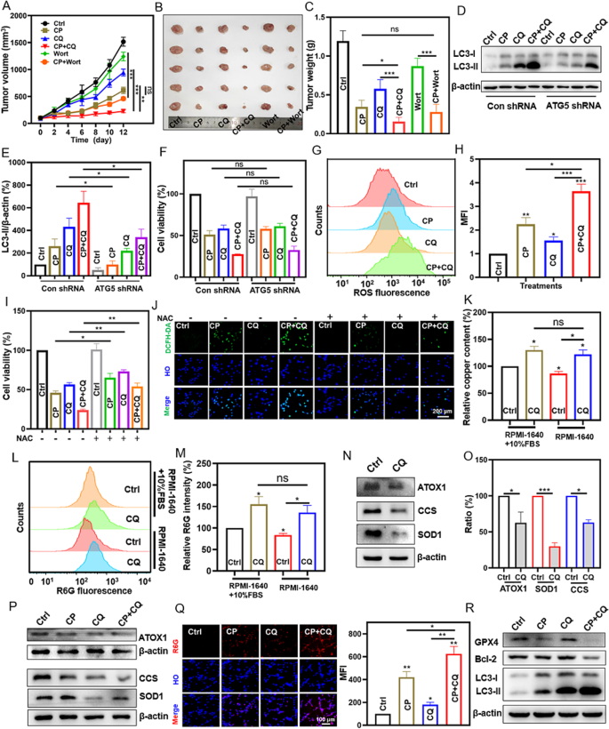
Synergistic tumor inhibition of CP and CQ depending on ROS technology and no matter autophagosome accumulation. (A) Tumor quantity curves have been monitored throughout completely different remedies by intratumoral injection. CP, 5 mg/kg; CQ, 8.56 mg/kg; wortmannin (Wort), 0.35 mg/kg. n = 5. (B and C) {Photograph} pictures and common tumor weights of the dissected tumors after the indicated remedies. n = 5. (D and E) Western blot evaluation (D) of CT-26 cells uncovered to the indicated remedies within the presence or absence of ATG5 expression attenuation. Picture J software program was used (E). CP, 15 µg/mL; CQ, 50 µM. Triple particular person experiments have been carried out. (F) Cell viability evaluation of CT-26 cells after the indicated remedies for twenty-four h within the presence or absence of ATG5 expression attenuation. CP, 15 µg/mL; CQ, 50 µM. Triple particular person experiments have been carried out. n = 3. (G and H) ROS evaluation by circulate cytometry after CT-26 after uncovered to completely different remedies for twenty-four h after which stained with DCFH-DA (10 µM; 20 min). Triple particular person experiments have been carried out. (I and J) Cell viability evaluation (24 h therapy) and ROS evaluation (6 h therapy) of CT-26 cells after the indicated remedies. CP, 15 µg/mL; CQ, 50 µM; NAC, 5 mM. (Ok) Whole copper contents in CT-26 cells after the indicated remedies for 12 h was quantified by ICP-OES. CQ, 50 µM. (L and M) Movement cytometry evaluation of R6G (10 µΜ; 20 min) staining in CT-26 cells after publicity to the completely different remedies for six h. CQ, 50 µΜ. Triple particular person experiments have been carried out. (N and O) Western blot evaluation of LC3 proteins in CT-26 cells uncovered to the remedies for 12 h and the outcomes have been analyzed by advantage of Picture J software program. CQ, 50 µM. The experiments have been repeated thrice. (P) Western blot evaluation of ATOX1, CCS and SOD1 proteins in CT-26 cells after the indicated remedies for 12 h. CP, 15 µg/mL; CQ, 50 µM. (Q) Fluorescence pictures stained by R6G (10 µΜ; 20 min) for CT-26 after the indicated remedies for six h. CP, 15 µg/mL; CQ, 50 µΜ. λex = 495 nm. (R) Western blot evaluation of CT-26 cells after the indicated remedies for twenty-four h. CP, 15 µg/mL; CQ, 50 µM
Immunogenic cell loss of life induction through reactive oxygen species technology
The above outcomes demonstrated a synergistic tumor inhibition of CP and CQ depending on ROS technology. On this examine, so we wandered whether or not the joint use of CP and CQ might induce immunogenic cell loss of life options in tumor cells. ICD is featured by elicitation of distinct DAMPs, together with publicity of CRT on cell membrane, HMGB1 secretion, and ATP launch. Herein, CP was demonstrated to set off ICD, nonetheless, upon combing with CQ, induced a strong and long-lasting ICD characteristic. CRT sub-cellular location was visualized by immunofluorescent staining. As confirmed in Fig. 4A, CRT was primarily expressed within the cytoplasm of CT26 tumor cells within the management and CQ-treated teams, nevertheless, an apparent CRT translocation to the cell membrane was noticed in CT-26 cells handled with the mixed administration of CP and CQ for 12 h, extra pronounced than the CP therapy group (Fig. 4A). After 24 h therapy, each the joint group and CP group exhibited outstanding CRT cell membrane localization (Fig. 4A). In the meantime, we detected the extracellular launch of HMGB1 within the cell cultured media by western blotting. The therapy with CP plus CQ for 12 h elicited extra important HMGB1 extracellular launch together with the decreased intracellular expression degree, whereas CP or CQ didn’t (Fig. 4B and S6a). After 24 h therapy, CP alone therapy really enhanced HMGB1 launch, however each the consultant western blot outcome and the statistical knowledge constantly advised that the joint use of CP and CQ did higher (Figs. 4B and S6b). The extra experiment was carried out to analyze the ATP launch after the completely different remedies. As proven in Determine S7, CP might considerably induce ATP launch into the cell tradition medium. Moreover, CQ alone wouldn’t considerably induce ATP launch, and affect CP-induced ATP launch. Collectively, these outcomes demonstrated that the joint use of CP and CQ might induce ICD associated options.
The ‘built-in stress response’ (ISR) is derived from the phosphorylation of eukaryotic translation initiation issue 2 subunit alpha (eIF2α) [6]. In comparison with cytotoxic medication that don’t induce ICD results, most strategies of inducing ICD results can successfully stimulate eIF2α phosphorylation, which is taken into account an necessary marker of ICD [7]. The unfolded protein response (UPR) triggered by endoplasmic reticulum (ER) stress is a part of the ISR. Throughout the activation of the UPR, IRE1α (a sensor on the ER membrane) cleaves XBP1 mRNA by way of its endoribonuclease exercise, producing the spliced type of XBP1 [64]. The reactive oxygen species (ROS) was prominently elevated after the mixed use of CP and CQ therapy. The ROS -mediated ER stress in pre-apoptotic stage cells is clearly linked to immunogenic cell loss of life (ICD). As proven in Fig. 4C and Determine S8, relative to CP or CQ -treated group, the joint use of CP and CQ triggered a extra important phosphorylation of eIF2α and notable expression of XBP1. Furthermore, the phosphorylation of eIF2α may very well be abolished by the ROS inhibitor NAC. Additional experiments have been carried out and demonstrated that the joint use of CP and CQ triggered immunogenic cell loss of life through ROS technology. Abrogation of ROS technology by an antioxidant agent (NAC) eradicated CP + CQ induced CT26 tumor immunogenic cell loss of life, as evidenced by the silence of CRT translocation to the cell membrane and HMGB1 extracellular launch (Fig. 4D and G). However, large accumulation of autophagosomes induced by the joint use of CP and CQ didn’t contribute to the synergistic tumor cytotoxicity (Fig. 3D and F), nevertheless, associated with ER stress induction, HMGB1 launch and the publicity of CRT on cell membrane (Determine S9a-b and S10a-S10c). To summarize, these biomarkers have been often called DAMPs and their concomitant presence implied immunogenic cell loss of life. Apart from, the joint use of CP and CQ elicited an early onset, above all, long-term high-level existence of ICD markers, which is strongly related to ROS technology and autophagosome accumulation.

ICD induced by the joint use of CP and CQ was associated to ROS-mediated ER stress. (A) CRT immunofluorescence imaging of CT-26 cells after uncovered to the indicated remedies for 12–24 h. (B) Western blot evaluation of HMGB1 within the supernatant (SN) and cytosol (CS) after CT-26 cells uncovered to the indicated remedies for 12–24 h. (C, E-G) CT-26 cells uncovered to the indicated remedies for 12 h have been subjected to western blot evaluation for phosphorylated eIF2α (p-eIF2α), HMGB1 in supernatant (SN) and cytosol (CS). The decrease panel of (C) confirmed the statistical knowledge. The statistical knowledge (F and G) was from (E). The experiments have been repeated thrice. CP, 15 µg/mL; CQ, 50 µM; NAC, 5 mM. (D) CRT immunofluorescence imaging of CT-26 cells after the indicated remedies for 12 h. CP, 15 µg/mL; CQ, 50 µM; NAC, 5 mM
CC@LPF exhibiting enhanced tumor cell uptake, selective tumor cytotoxicity, and biosafety
As illustrated above, the joint use of CP and CQ, not solely realized the maximized direct killing impact on tumor cells but additionally elevated the immunogenicity of the native tumor micro-environment, was thought-about as an efficient technique for tumor therapy. Nonetheless, there are nonetheless sure limitations to copper peroxide or chloroquine primarily based tumor therapy strategies, such because the potential systemic toxicity, low effectivity of tumor concentrating on, and so forth. [3, 65]. Herein, to extend the effectivity of CP in tumor suppression mixed with CQ, Folic acid (FA) conjugated and PEGylated liposomes (LFA) have been employed for the supply of CP and CQ (Determine S11). The as-synthesized FA conjugated PEGylated liposomes carrying CP and CQ (denoted as CC@LPF; Fig. 5A and S11) obtained systematic characterization. The morphology of the as-prepared CC@LPF hardly modified in comparison with CP nanospindles (Fig. 5A). The zeta potential of CQ@CP was ~-10 mV, whereas that of CP nanospindles was ~ 5.02 mV, implying the feasibility of loading CQ on CP nanospindles through electrostatic adsorption (Fig. 5B). Then the negatively charged CQ loaded CP nanospindles (CQ@CP) have been adorned by folate conjugated DSPE-PEG (denoted as CC@LPF) through hydrophilic-hydrophobic interactions. The CC@LPF nanoparticles offered a zeta potential of ~-21.33 mV, which was useful for circulation stability of the in vivo therapy. As well as, the dimensions of CC@LPF elevated to ~ 276.1 nm after the modification of folate conjugated DSPE-PEG2000-FA (Fig. 5C). We additionally investigated the colloidal stability of CC@LPF nanoparticles. In every of the options, together with PBS, 0.9% saline, RPMI-1640 tradition medium and 10% fetal bovine serum, the PDI of CC@LPF was all the time beneath 0.20 (Determine S12a-S12d), suggesting a great dispersibility. Apart from, CC@LPF, even maintained in numerous options at room temperature for 14 days, additionally exhibited no important adjustments on the dimensions and PDI (Determine S12a-S12d), indicating a superb colloidal stability. Within the FTIR spectra, the bands positioned at 1 680 cm− 1 and 1 050 cm− 1 have been assigned to the stretching vibration of C = O bonds and the asymmetrical stretching vibrations of -C-O-C- of DSPE-PEG2000-FA, respectively, suggesting the profitable PEGylation of CQ@CP (Fig. 5D). As well as, the band of the CC@LPF positioned at 1 611 cm− 1 was attributed to the bending vibration of N-H bonds of CQ, suggesting the profitable loading of CQ on CP nanospindles. In the meantime, the UV-Vis spectra of CQ@CP and CC@LPF demonstrated one attribute absorption peak at 343 nm of CQ whereas CP@LPF offered no absorption peaks, additional illustrating the profitable carrying of CQ (Fig. 5E). The optimized encapsulation and loading effectivity of CQ have been calculated to be 44.48% and 17.37%, respectively. As well as, the encapsulation and loading effectivity of CP have been 92.67% and 9.04%, respectively.
Then the copper ion launch efficiency of CC@LPF at varied pH values was investigated utilizing ICP-OES. Roughly, copper ions burst out from CC@LPF after incubation at pH 5.5 for 1 h (Fig. 5F). After 24 h, about 67.81% copper ions liberated from CC@LPF, which probably facilitated the GSH consumption and ROS manufacturing for CDT therapeutic efficacy. In distinction, copper ions have been scarcely launched within the impartial medium, implying a superb pH-responsive launch efficiency of CC@LPF. The technology of ∙OH by CC@LPF was examined utilizing MB and three,3’,5,5’-tetramethylbenzidine (TMB) as colorimetric probes. Inspiringly, CC@LPF induced an identical degradative impact of MB in comparison with CP nanospindles on the similar pH values and concentrations, indicating the modification of CQ and FA didn’t weaken the ROS manufacturing of CP nanospindles (Fig. 5G). Furthermore, CC@LPF and CP induced related TMB chromogenic response on the similar situation, additional confirming the ROS technology of CC@LPF (Determine S13). Folic acid (FA) receptors are extremely expressed on the mobile membranal floor of varied most cancers cells. Accordingly, FA was broadly used as an energetic, protected, and environment friendly concentrating on molecule to most cancers cells. Herein, the concentrating on effectivity of CC@LPF was investigated by detecting the variation of intracellular copper focus. As proven in (Determine S14a), compared with CP nanospindles, the intracellular copper focus elevated to ~ 1.76 fold after incubation with CP@LPF for six h, indicating the remarkably enhanced uptake of CC@LPF.
To additional uncover the mechanism of FA enhanced cell uptake, free FA was used to dam the FA receptor on the floor of most cancers cell, and FA conjugated and PEGylated liposomes carrying FITC, CP and CQ (denoted as FITC/CC@LPF) have been constructed (Determine S14b). After the incubation with FITC/CC@LPF for the completely different time, CT-26 cells exhibited time-dependent improve of FITC fluorescence depth (Determine S14c). After incubation with FITC/CC@LPF for two h and 6 h, the confocal laser scanning microscope pictures offered vivid inexperienced fluorescent sign (Fig. 5H). In distinction, nearly no inexperienced fluorescence offered when incubated with free FA and CC@LPF (Fig. 5I), suggesting that FA performed an necessary function on cell uptake of CC@LPF nanoparticles. Taken collectively, the profitable loading of CQ and conjugation of FA of CC@LPF served as a H2O2 self-supplied, pH-responsive and energetic concentrating on nanoplatform for CDT tumor remedy.

Preparation of CC@LPF for enhanced tumor cell uptake. (A) TEM pictures of CC@LPF. (B) Zeta potentials of varied nano-formulations. (C) DLS measurement distributions of varied formulations. (D) FTIR spectra of CP, CQ, LPF, CP@LPF, CQ@LPF, CC@LPF. (E) The UV–vis absorption spectra of CP, CQ, CQ@CP, LPF, CP@LPF, CQ@LPF, CC@LPF. (F) pH responsive launch conduct of CC@LPF within the completely different pH conditioned PBS. (G) UV-Vis spectra demonstrating the degradation of MB by CP nanospindles and CC@LPF with an equal focus (50 µg/mL) underneath varied pH values. (H and I) Fluorescence imaging for the uptake of FITC/CC@LPF CT-26 cells after 2 h and 6 h incubation within the presence of folic acid (1 mM) or not. The experiment was repeated thrice
As a result of particular mixture of folate with folate receptor overexpressed on the membranal floor of tumor cells, CC@LPF may very well be extra successfully endocytosed, and exhibited extra important ROS technology plus tumor cell toxicity. As proven in Fig. 6A, CC@LPF precipitated extra apparent ROS technology, relative to the joint use of CP and CQ, CP@LPF or CQ@LPF. MTT assay was additional carried out and indicated that CC@LPF therapy triggered extra lower of cell viability relative to both CP@LPF or CQ@LPF therapy (Fig. 6B). Moreover, CC@LPF therapy elicited ~ 82.65% cell viability loss, greater than that of the joint use of CP and CQ (Fig. 6B). Calcein AM, a hydrophobic and cell permeable dwelling cell dye, upon getting into the cells, could be hydrolyzed to calcein by intracellular esterases and emit inexperienced fluorescence after the mix of calcein and intracellular calcium ions. Herein, fluorescence pictures clearly confirmed that CC@LPF gave rise to probably the most lower of Calcein AM staining, and in line with which, the utmost cell loss of life (PI positve) was additionally showing (Fig. 6C). These outcomes have been additionally confirmed by a further statistical knowledge of the ratio of reside cells/whole cells (Fig. 6D). Moreover, apoptosis evaluation was additionally carried out. As confirmed in Fig. 6E, folate conjugated lipid system might additional reinforce tumor cell apoptosis induction, and CC@LPF exhibited probably the most apoptosis elicitation.
The liposome supply technique couldn’t improve the renal clearance price of CP, however might enhance the blood compatibility of CP. We evaluated the renal clearance time price of CP and CC@LPF. As proven in Determine S15, the renal clearance price of CP and CC@LPF nanoparticles was much less then 7% at 96 h publish injection, suggesting their low renal clearance effectivity. The low renal clearance price was in all probability as a result of measurement limitations of glomerular filtration, which allowed the excretion of nanomaterials with the dimensions beneath 6 ~ 8 nm [66]. The low renal clearance effectivity of CC@LPF was probably favorable for the in vivo therapeutic efficacy, nonetheless, which might additionally convey issues about systemic toxicity. Therefore, some crucial security evaluations are carried out. Hemolysis evaluation was first carried out and evidenced that the as-synthesized CC@LPF nanocomposite possess blood compatibility. As proven in Fig. 6F, each CP and CP + CQ therapy teams offered important hemolysis, whereas CC@LPF therapy group didn’t. Tumor concentrating on liposomal supply of CP and CQ offered selective tumor cell development inhibition and circumvented CP induced hemolysis. Herein, three simply out there non-cancerous cell strains, together with human embryonic kidney 293T (HEK-293T), Reworked Human Liver Epithelial-3 (THLE-3) and L929 (L-929) mouse fibroblast, have been deployed for the comparative investigation. As exhibited in Fig. 6G, CC@LPF provoked a most development inhibition in CT26 cancerous cells relative to a different three non-cancerous cell strains. Therefore, Tumor concentrating on liposomal supply of CP and CQ improved tumor cell selectivity and blood compatibility of mixture remedy.

CC@LPF exhibited enhanced and selective tumor cytotoxicity with out inflicting hemolysis. (A) Fluorescence pictures of CT-26 cells dyed with DCFH-DA probe after completely different remedies for six h. Automobile management: Ctrl. (B) Cell viability evaluation of CT-26 cells uncovered to the completely different remedies for twenty-four h. The experiment was repeated thrice. n = 3. (C and D) Calcein AM and propidium iodide (PI) staining of CT26 cells uncovered to the completely different remedies for twenty-four h. The corresponding statistical evaluation have been carried out by Picture J software program. The ratios of Dwell/whole cells have been indicated by Calcein AM (2 µM) and PI (5 µM) staining for 25 min. n = 3. (E) Apoptosis evaluation of CT-26 cells uncovered to the completely different remedies for twenty-four h. (F) Hemolysis experiments of various concentrations (equal to copper ions) of CP, CQ, CP + CQ, CC@LPF. The unit of the numbers within the determine is μg/mL. (G) Cell viability evaluation of CT-26, 293T, THLE-3 and L929 cells after the therapy with CC@LPF at completely different concentrations for twenty-four h. The experiment was repeated thrice (n = 3). The concentrations used of all teams have been equal to that of copper ions
In vivo tumor concentrating on and anti-tumor efficacy of CC@LPF with a PD-L1 inhibitor or not
In vivo tumor concentrating on supply of CC@LPF was additional studied by recording the NIR fluorescence of ICG, which was at the moment authorised by the US Meals and Drug Administration (FDA) as a NIR imaging reagent for scientific use. In the meantime, to proof FA performed an necessary function in energetic focused supply functionality, FA conjugated and PEGylated liposomes carrying ICG, CP and CQ (denoted as ICG/CC@LPF) and PEGylated liposomes carrying ICG, CP and CQ (denoted as ICG/CC@LP) have been ready. Fluorescence emission spectra and the corresponding fluorescence pictures of ICG/CC@LPF and ICG/CC@LP with an equal ICG focus demonstrated that ICG was efficiently encapsulated in liposomal system (Determine S16a-b). The fluorescence emission spectra confirmed the emission wavelength round 820 nm. DLS outcomes demonstrated the imply measurement of ICG/CC@LPF was round 266.93 nm, bigger than ICG/CC@LP, indicating the FA coating (Determine S16c). The true-time NIR imaging of mice bearing CT-26 cell transplantation tumors after tail vein injection of ICG/CC@LPF or ICG/CC@LP was additional monitored. As proven Fig. 7A and B, we might see that the ICG/CC@LPF was transported into tumor tissue after 0.5 h and reached the utmost depth at 12-hour time-point, then the sign starting to fade. Apart from, ICG/CC@LPF exhibited extra accumulation at tumor websites relative to ICG/CC@LP (Fig. 7B). By advantage of the ex vivo fluorescent imaging of tumors and organs collected at 12 h publish injection, it may very well be clearly captured that ICG/CC@LPF offered the strongest NIR fluorescent indicators, greater than these of ICG/CC@LP group (Fig. 7C), demonstrating folic acid enhanced tumor concentrating on supply.

In vivo tumor concentrating on and anti-tumor efficacy of CC@LPF with or with out α-PDL1. (A and B) In vivo NIR imaging of tumors at completely different time factors after tail injection of ICG/CC@LP and ICG/CC@LPF (1 mg/kg ICG). The tumors have been circled and relative fluorescence depth of tumors was analyzed by Picture J software program. (C) Ex vivo imaging at 12 h post-injection. (D) A schematic illustration for the therapeutic mannequin. Intraperitoneal injection (i.p.) for α-PDL1; intravenous injection (i.v.) for CC@LPF and others. (E and F) CT-26 xenograft tumor development curves after tail injection with completely different remedies. Saline: physiological saline. α-PDL1, 2 mg/kg. The concentrations of all different therapy group have been equal to that of copper ions 5 mg/kg, and the dose of CQ was calculated as 9.60 mg/kg. (G) {Photograph} pictures of dissected tumors from the completely different teams. (H) Tumor inhibition price of various teams. n = 5. (I) Physique weight variations of mice
Anti-tumor effectivity of CC@LPF and additional joint therapy of CC@LPF and PD-L1 blockade antibody have been investigated. Herein, we additionally noticed the mixed anti-tumor impact of CC@LPF through intravenous injection. A schematic illustration for the therapeutic mannequin was exhibited in Fig. 7D. As proven in Fig. 7E and H, CC@LPF exhibited the strongest tumor inhibition impact relative to both of CP@LPF or CQ@LPF, as evidenced by probably the most important lower of tumor quantity and tumor weight, underlying the effectiveness of the mixed therapy. ICD refers to an immune stimulated cell loss of life mode in most cancers therapy, which is a promising technique for sensitizing PD-1/L1 immune checkpoint blockade (ICB) remedy. Contemplating the CC@LPF might induce selective tumor cell toxicity and ICD, we investigated the in vivo anti-tumor efficacy and security of the joint therapy between CC@LPF and PD-L1 blockade antibody. For this goal, we first evaluated the expression of PD-L1 in CT-26 by western blotting. As displayed in Determine S17, CC@LPF wouldn’t lower the basal expression of PD-L1, underlying the potential for the joint use of CC@LPF and ICB for tumor remedy. Subsequent, CT26-bearing BALB/c mice accepted the completely different drug formulations, and the tumor inhibition effectivity of various remedies have been monitored. Herein, the dosage of PD-L1 blockade antibodies was referring to the earlier literature [67]. As proven in Fig. 7F and H, in line with the earlier research [67], PD-L1 ICB monotherapy was poorly responsed in CT26 tumor mannequin, nevertheless, PD-L1 ICB antibody might considerably improve the antitumor impact of CC@LPF.
The completely different therapeutic teams exhibited negligible systemic toxicity. As illustrated above, CC@LPF exhibited each selective tumor cell toxicity and blood compatibility. Apart from, the animal physique weights within the completely different teams have been additionally monitored throughout the remedy and the outcomes indicated that completely different therapeutic fashions wouldn’t elicit mouse physique weight reduction (Fig. 7I). Moreover, contemplating the present liver distribution conduct (Fig. 7C) and low renal clearance price of CC@LPF (Determine S15), some blood biochemical indexes associated to liver or kidney harm, have been additional monitored to proof the security of CC@LPF primarily based therapeutic technique. As proven in Determine S18a-S18d, the completely different remedies in mice, together with CC@LPF, wouldn’t clearly have an effect on the extent of alkaline phosphatase, alanine aminotransferase, aspartate aminotransferase or blood urea nitrogen, suggesting the security of CC@LPF primarily based therapeutic technique.
Collectively, the as-synthesized CC@LPF, potentiated as tumor remedy brokers, exhibited a negligible systemic toxicity and the capabilities of tumor cell killing and synergistic tumor remedy with ICB.
The mechanism of the joint anti-tumor impact of CC@LPF and ICB
Then, the mechanism of the joint therapy of CC@LPF and PD-L1 blockade antibody was investigated on the organizational and mobile ranges. The direct influence of ICD in tumor microenvironment acts on the dendritic cells (DCs). The launched HMGB1 and CRT from tumor cells bind TLR4 and CD91 on the DCs, respectively, ensuing within the DCs mature and IL-1β secretion. Furthermore, the tumor antigen was internalized and offered by DCs, which might result in the activation of tumor infiltrating T cells. Right here, we firstly detected the CD11c+ MHC-II+ DCs infiltration in tumor microenvironment (TME) by circulate cytometry in accordance with the gating technique in Determine S19. As proven in Fig. 8A and C, the proportion of CD11c+ MHC-II+ DCs cells have been clearly raised in CC@LPF therapy group. Intriguingly, CQ@LPF, however not CP@LPF, considerably elevated the proportion of CD11c+ MHC-II+ DCs, and there was no important distinction between CC@LPF and CQ@LPF, suggesting CC@LPF regulated the proportion of CD11c+ MHC-II+ DCs primarily through CQ. These outcomes have been additionally in line with the earlier reviews, which indicated that CQ might abrogate lysosome-dependent degradation of MHC-II [68, 69] and promote DCs mature plus antigen presentation [70, 71]. Furthermore, α-PDL1 monotherapy offered no affect on the proportion of DCs on this analysis mannequin. Apart from, though the mix of α-PDL1 + CC@LPF additional elevated the proportion of DCs in TME, there was no important distinction between α-PDL1 + CC@LPF and CC@LPF therapy teams (Fig. 8A and C), which indicated that the CC@LPF mono-treatment might effectively increase the DCs infiltration within the TME. We additional detected the T cell infiltration within the TME by IHC staining and circulate cytometric evaluation. Herein, the circulate cytometric evaluation for CD3+ T cells and CD3+ CD8+ T cells have been in accordance with the gating methods in Determine S20 and S21. Persistently, in comparison with these of CC@LPF therapy group, tumors grown on the mice uncovered to CC@LPF plus α-PDL1 antibodies confirmed considerably improved proportions of T cells (CD3+) and cytotoxic T cells (CD3+CD8+) (Fig. 8B, D and E, S22a-S22b), which revealed that CC@LPF might sensitize α-PDL1 remedy. Apart from, the infiltration of cytotoxic CD8+ T cell considerably elevated within the CC@LPF group in comparison with CP or CQ mono-treatment. Thus, CC@LPF was a promising candidate to spice up the tumor immunotherapy.
Ki-67, outlined as a nuclear protein primarily associated to proliferating cells, and functionally associated to cell mitosis, can mirror the exercise degree of tumor cell proliferation. IHC staining outcomes demonstrated that Ki67 expression in CC@LPF therapy group considerably decreased in comparison with both CP@LPF or CQ@LPF therapy group, additional suggesting the benefit of the joint use of CP and CQ. PD-L1 ICB antibody, α-PDL1, alone presenting no decline of Ki67 expression, however would markedly reinforce CC@LPF elicited Ki67 inhibition (Fig. 8F and G). Apart from, the morphology of organ and tumor tissue sections in seven teams of mouse fashions was studied by way of H&E staining (Fig. 8H and S23). It was clearly displayed nuclear atypia and inferior staining of chromatin for CC@LPF therapy relative to both CP@LPF or CQ@LPF therapy, although each of which confirmed sure diploma of anti-tumor impact. α-PDL1, alone presenting no apparent histological change, nevertheless, CC@LPF + α-PDL1 resulted in additional important nuclear atypia and inferior staining relative to CC@LPF therapy alone (Fig. 8H). The adjustments in cell integrity and tissue morphology of the primary organs in numerous therapy teams may very well be negligible (Fig. 8H and S23). These outcomes demonstrated that CC@LPF potentiated as tumor remedy brokers exhibiting the capabilities of tumor cell killing and synergistic tumor remedy with ICB.

The mechanism of the in vivo joint anti-tumor effectivity of CC@LPF and ICB. (A) Movement cytometric evaluation of the populations of CD11C+ MHC-II+ DCs in tumor setting after wouldn’t clearly have an effect on the extent. (B) CD3, CD8 IHC staining of tumor tissue sections obtained from tumor-bearing mice after varied remedies. H, hematoxylin. (C) The chances of MHC-II+ DCs from CD11c+ DCs in tumor microenvironment. n ≥ 4. (D-E) Movement cytometric evaluation of the populations of CD3+ and CD3+ CD8+ T cells in TME after the completely different remedies. n ≥ 4 (F) Quantification of Ki67 optimistic space in tumors. n = 3. The corresponding statistical evaluation have been carried out by Picture J software program. (G) Ki-67 IHC staining of tumor tissue sections obtained from tumor-bearing mice with varied remedies. H, hematoxylin. (H) Pathological evaluation of the very important organs together with kidney, lung, liver and tumor gathered from CT26 tumor loaded mice after completely different remedies by H&E staining