Comparability of immunophenotype between 2- and 16-month murine fashions
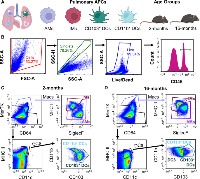
We first sought to characterize the basal APC phenotype throughout the 2- and 16-month C57BL/6 mice. We established a flow-cytometry gating scheme of single-cell complete lung digests to determine AMs, IMs, CD103+ DCs, and CD11b+ DCs within the two age teams (Fig. 1A, staining info Supplemental Desk S1) [25]. After gating for cells, adopted by singlets, after which the dwell cell inhabitants, the samples have been additional remoted for myeloid origin by means of CD45+ expression (Fig. 1B). Following this, macrophages could possibly be remoted by excessive CD64 and MerTK expression, which have been then additional categorized as AM or IM utilizing SiglecF and CD11b expression. DCs have been remoted from their low CD64 and MerTK however excessive MHCII and CD11c expression. These have been then categorized as CD103+ or CD11b+. Nevertheless, when making use of the identical gating scheme to the aged mannequin (16-months), there seemed to be appreciable shifts within the markers used for identification of cell sorts. Macrophages exhibited larger MerTK expression general and a drastic enhance in CD11b expression for the AM inhabitants (Fig. 1D). Whereas the DC gate confirmed comparable expression of MHC II and CD11c in comparison with the younger group, the ungated portion of the pattern confirmed a rise in MHC II general, considerably hindering the analogous gating scheme for DCs from the younger mice (Fig. 1D). Moreover, the aged mannequin confirmed a noticeable lower in CD103+ DC occasions, and the introduction of a brand new cell kind that was neither CD103+ or CD11b+, labeled as DC3 (Fig. 1D). This DC3 group confirmed noticeably low expression of CD11b, CD45, and MHC II compared to the opposite DC sorts (Supplemental Determine S2) however was a novel and separate inhabitants from both of the opposite DC sorts, as proven by means of again gating methods (Supplemental Determine S2). Earlier literature has not explored shifts in lung DC populations in growing old research, however as these cell populations on this age group particularly have not often earlier than been studied, it’s not stunning that distinctive phenotypes could also be found.
Superior age demonstrates shift in APC inhabitants phenotype. Entire murine lung samples have been digested right into a single cell suspension for multicolor stream cytometry evaluation of 4 pulmonary antigen presenting cells (APCs) of curiosity (alveolar macrophages (macs.; AMs), interstitial macrophages (IMs), CD103+ dendritic cell (DCs), and CD11b+ DCs throughout two ages (2- and 16-months). (A) Examine overview schematic together with cells and age fashions of curiosity. (B) Main gating for cells, singlets, dwell cells, and CD45+ cells. C–D.) Consultant stream gating scheme for 2-months (C) and 16-months (D)
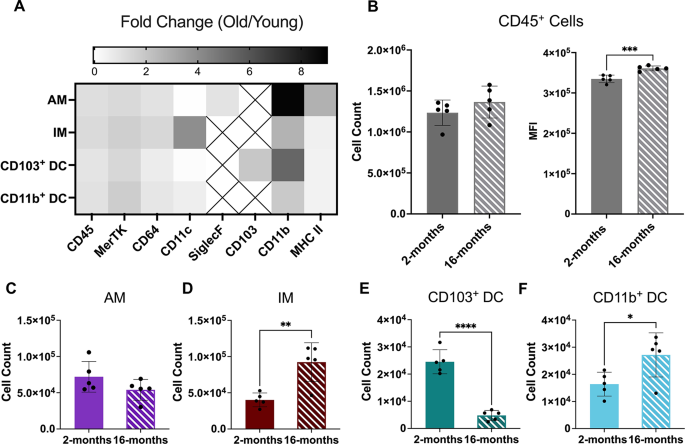
Inhabitants phenotype expression comparability between the 2 ages confirmed comparable tendencies in relative expression for the 8 markers chosen (CD45, MerTK, CD64, CD11c, SiglecF, CD103, CD11b, and MHC II; discuss with Supplemental Determine S3). Nevertheless, obvious shifts in expression of sure markers have been noticeable between the 2 teams. Thus, relative fold change for every marker of the untreated 16-month group was in comparison with untreated younger mice to characterize adjustments in phenotype of the 4 APCs (Fig. 2A). Right here, essentially the most vital change noticed for all cell sorts was a rise in CD11b expression, with a virtually 10-fold enhance for AMs and 6-fold enhance for CD103+ DCs (Fig. 2A). AMs additionally demonstrated a big enhance in MHC II expression in comparison with different cell sorts (Fig. 2A). These outcomes for AMs are consistent with different beforehand reported evaluation of aged lung fashions, however as of but had not been quantified in a pre-senescent, 16-month aged mannequin [47]. One other attention-grabbing outcome was a considerable enhance in CD11c expression for the IMs (Fig. 2A). This cell kind doesn’t sometimes current CD11c; nonetheless, different research have discovered higher presence of CD11c+ macrophages in infected microenvironments and it’s identified that this marker is inherently concerned in phagocytosis [48,49,50]. Additional quantitative evaluation from untreated information confirmed a slight but nonsignificant enhance in CD45+ cell counts however an general vital enhance in CD45 expression within the 16-month age group (Fig. 2B). Consultant frozen lung histology additional confirmed that CD45 expression elevated on the tissue-level utilizing a major CD45 IHC stain (discuss with Supplemental Determine S4 A-D).
16-month lung presents distinctive mobile immunophenotype. Mobile comparability was carried out of the murine lung for 2 age teams of mice (2- and 16-months). (A) Fold change in floor marker MFI of 16-month remoted cell sorts in comparison with common expression of the 2-month group. Packing containers with Xs point out markers that weren’t expressed in these cell sorts. (B) Multicolor stream cytometry evaluation of general CD45+ cell counts and CD45 median fluorescence depth (MFI). C-F.) Circulate cytometry remoted particular person cell counts for alveolar macrophages (AM; C), interstitial macrophages (IM; D), CD103+ dendritic cells (DCs; E), and CD11b+ DCs (F) within the two ages. Displayed numerical outcomes represents imply ± SD (n = 5) from untreated group outcomes. Indicated significance is calculated by way of unpaired Pupil’s t-tests [p < 0.05 (*), 0.01 (**), 0.001 (***), < 0.001 (****)]
Furthermore, cell counts for the 4 APC cells of curiosity all demonstrated adjustments with age (Fig. 2C-F). Right here, the 16-month age demonstrated noticeable but nonsignificant lower in AM cell counts accompanied with a complementary vital enhance in IM cell counts (Fig. 2C and D). This phenomenon has been noticed beforehand in literature and is believed to be the results of a turnover of guard from AM-dominated clearance in younger people to a extra CD11b+ cell kind dominated lung inhabitants over time in aged lungs [51, 52]. Apparently, the DC cells additionally confirmed an analogous development, through which there was a discount in CD103+ DCs occasions, beforehand seen in Fig. 1D, accompanied by a rise in CD11b+ DC rely, which has but to be reported in literature (Fig. 2E and F). Further consultant H&E staining of frozen lung tissue demonstrated no vital inflow of inflammatory our bodies within the 16-month pattern in comparison with the 2-month mice (discuss with Supplemental Figures S4E-F).
Altered in vivo nanoparticle uptake by aged APCs within the lung
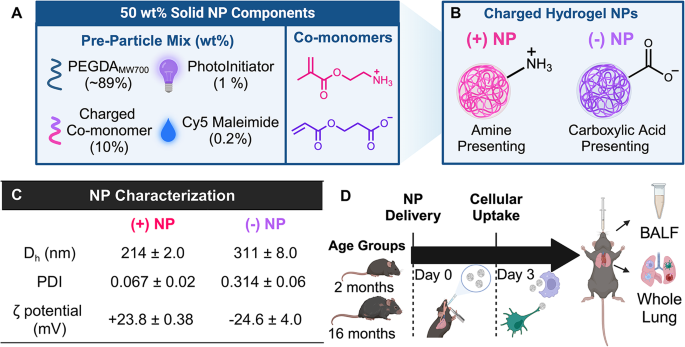
To be able to quantify the change in NP uptake of those distinctive age teams, an inert PEGDA hydrogel NP was synthesized utilizing a reverse emulsion photopolymerization approach. This particle chemistry has broadly been utilized in literature, because the monomeric elements are readily modifiable for tunability of design (Fig. 3A) [38, 42, 44, 45, 53]. Moreover, comparable PEGDA NPs synthesized with the identical cationic and anionic monomeric elements in single-dose, quick time period research have proven non-inflammatory responses within the lung, making them perfect autos for pulmonary phagocytic research of APCs with out induction of a pro-inflammatory impact [38, 53]. Right here, totally different NPs cost was evaluated to find out preferential uptake between the aforementioned APCs of curiosity reported in younger and previous mice. Two formulations of PEGDA NPs have been synthesized with opposing floor expenses by inclusion of two charged useful monomers (Fig. 3A). The cationic charged useful monomer contained an amine-presenting useful group, which might be known as (+)NP and proven in shiny pink information figures for the rest of this text (Fig. 3B). The anionic monomer contained a carboxylic acid-presenting group, known as (-)NP and proven as shiny purple information figures from hereon (Fig. 3B). The NPs synthesized right here displayed excessive uniformity in hydrodynamic dimension (Dh), polydispersity index (PDI), and zeta potential (Fig. 3C). Each formulations achieved NPs round 200–300 nm in Dh and an absolute zeta potential round 25 mV (Fig. 3C). Right here, it was desired to attain zeta potentials higher than 20 mV to make sure distinctions in NP cost and adequate electrostatic interactions between NPs to attenuate aggregation over time. Whereas there may be roughly 100 nm distinction in Dh of the 2 formulations, the (-)NP had a bigger PDI pointing to some extent of polydispersity to make sure an inexpensive overlap in particle sizes, such that each formulations must be anticipated to be internalized by way of phagocytosis. Cryo-scattering electron microscopy photographs of hydrogel NPs additionally demonstrated excessive sphericity of the particles (discuss with Supplemental Determine S5). All additional research delivered these PEGDA NPs on to murine lungs by way of orotracheal instillation, a pulmonary supply approach that has proven restricted lung localization [38], and after a specified period of time, the BALF and complete lung have been efficiently acquired for evaluation (Fig. 3D). We selected to check NP uptake to an untreated group in each ages fairly than a PBS-only, placebo group since earlier research utilizing PBS-only teams haven’t demonstrated elevated inflammatory alerts or soluble elements and thus the untreated group serves as a helpful reference for NP distribution [43, 44, 54, 55].
PEGDA Nanoparticle Formulation and Dosing Timeline. Poly(ethylene-glycol) diacrylate (PEGDA) hydrogel nanoparticles (NPs) at 50 wt% stable have been synthesized utilizing a reverse emulsion photopolymerization approach for uptake research. (A) Composition of PEGDA pre-particle composition (left) and particular co-monomer chemical buildings (proper) of PEGDA NPs. (B) Floor presentation of synthesized NPs (pink = (+) NP, purple = (-) NP) (C) Dynamic gentle scattering information for NPs. Outcomes characterize imply ± SD, n = 2 unbiased batch replicates. Dh = hydrodynamic dimension, PDI = polydispersity index (D) Timeline of dosage experiments with NPs resulting in assortment of bronchoalveolar lavage fluid (BALF), serum, and complete lung tissue for evaluation
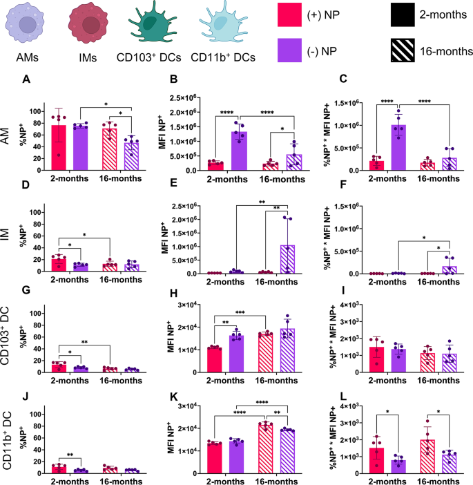
A preliminary examine delivering PEGDA NPs of each expenses by way of orotracheal instillation to 2-month-old murine lungs was used to evaluate short-term NP uptake after 24 h by way of %NP+(discuss with Supplemental Figures S6 and S7). Whereas there was vital uptake of each formulations by the macrophage populations, there was minimal uptake (lower than 10%) by DC populations (discuss with Supplemental Determine S6). Thus, the rest of the research carried out used an incubation interval of 72 h to permit for acceptable mobile uptake by all populations and to match beforehand reported literature (Fig. 3C) [38, 54]. From digested lung samples that have been handled with both formulation, NP uptake was quantified utilizing %NP+(Fig. 4A, D, G and J) in addition to median fluorescence depth (MFI), a mean fluorescence worth used to roughly decide the amount of NPs phagocytosed by NP+ cells (Fig. 4B, E, H and Okay). Moreover, a ultimate parameter of whole fluorescence that mixed these two beforehand talked about by means of multiplication was additional used to common NP uptake throughout these 4 cells of curiosity (Fig. 4C, F and I, & 4L).
Nanoparticle uptake shifts with cost, age, and kind of cell. Two ages of mice (2- and 16- months) have been dosed by way of orotracheal instillation with 100 µg of optimistic [(+)NP] or adverse [(-)NP] PEGDA NPs. After 72 h, stream cytometry decided NP uptake in 4 APCs within the lung together with alveolar macrophages (AMs, A–C), interstitial macrophages (IMs, D–F), CD103+ dendritic cells (DCs, G–I), and CD11b+ DCs (J–L). Parameters proven embrace % of cell inhabitants with NP uptake (%NP; A, D, G, J), median fluorescence depth of the NP channel (MFI; B, E, H, Okay), and the full fluorescence (%NP*MFI; C, F, I, L). Knowledge represents imply ± SD (n = 5). Indicated significance is calculated by way of two-way ANOVA [Tukey Test, p < 0.05 (*), 0.01 (**), 0.001 (***), < 0.001 (****)]
Of observe, AMs confirmed a considerably larger proportion of NP+ cells in comparison with some other group no matter age or floor cost (Fig. 4A). That is per identified research of AMs, which have proven that these AMs are the dominating phagocytic cell within the lung [20]. Within the 2-month group, the AM inhabitants confirmed practically 80% uptake after 72 h with no desire to both formulation primarily based on NP+ cells (Fig. 4A). Nevertheless, the aged group confirmed vital discount NP uptake, with a discount between the (-)NP uptake and (+)NP compared to their respective younger group controls (Fig. 4A). Apparently, all younger teams aside from AMs confirmed a desire for (+)NP uptake, with IMs, CD103+ DCs, CD11b+ DCs all exhibiting roughly 20% uptake after 72 h of (+)NPs (Fig. 4D, G and J). Apart from the CD11b+ DC group, the interstitial populations additionally demonstrated a big lower in (+)NP uptake within the aged group however no change within the (-)NP uptake (Fig. 4D, G and J).
Whereas the %NP+ uptake exhibited solely minor adjustments throughout age, extra noticeable outcomes emerged for NP+ MFI of the 4 APCs of curiosity. AMs of each age teams confirmed excessive preferential uptake for (-)NPs; nonetheless, the aged AM group demonstrated a extremely vital (p < 0.001) lower in uptake of (-)NPs (Fig. 4B). For the (+)NP, AMs confirmed no change in uptake throughout age (Fig. 4B). Moreover, whereas the IM inhabitants confirmed minimal uptake of NPs within the younger inhabitants in comparison with the AMs, there was a dramatic enhance (p < 0.01) within the amount of (-)NP uptake in NP+ cells within the aged inhabitants (Fig. 4E). There was noticeably higher variability throughout the IM inhabitants by way of (-)NP uptake; this will converse to how undefined and nonlinear the growing old course of will be between topics. Nevertheless, these outcomes are consistent with the idea that the IM inhabitants might tackle a extra phagocytic function within the lung with the declining operate of the AM inhabitants in aged people [20, 23]. Moreover, the mix %NP+*MFI for the AM and IM populations helps notion that anionic drug formulations have higher uptake by macrophages within the mucosa (Fig. 4C and F).
Compared to the dramatic change in uptake of NPs by macrophages, the DC populations general confirmed minimal uptake of NPs with general MFIs roughly 100 instances lower than that of the AM inhabitants (Fig. 4H and Okay). Whereas each DC sorts appeared to have a slight development towards (+)NP desire in %NP+, there was considerably larger amount of (-)NP uptake within the younger inhabitants of CD103+ DCs (Fig. 4H). Apparently, there seemed to be a rise in NP uptake for all formulations and DC sorts within the aged group, with CD103+ DCs rising in (+)NP uptake and CD11b+ DCs having a drastic enhance (p < 0.01) in each NP formulations (Fig. 4H and Okay). Thus, together with the %NP+ information, each DC populations confirmed no constant change in NP uptake with age, whereas solely the CD11b+ DC group exhibited preferential (+)NP uptake in each ages (Fig. 4I and L). Moreover, the opposite DC inhabitants remoted, DC3, was analyzed for NP uptake, which confirmed very comparable outcomes to the CD11b+ DC inhabitants (discuss with Supplemental Determine S8). Surprisingly, the DC3 subset confirmed elevated proportion of NP uptake however decreased MFI for NP+ cells in comparison with different DC sorts (discuss with Supplemental Determine S8A-C).
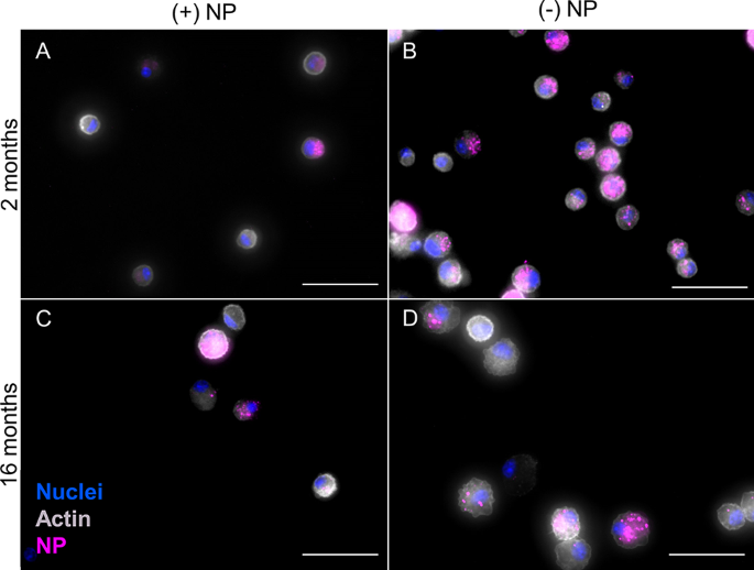
Along with complete lung stream cytometry outcomes, cells have been remoted from bronchoalveolar lavage fluid (BALF) to additional visualize adjustments in NP uptake throughout age. Thus, cells from BALF have been appropriately plated and handled to visualise nuclei (DAPI; blue), actin (Phalloidin; gray), and NPs (pink); Fig. 5). Right here, (+)NPs present comparable constant uptake throughout cells that’s comparable for each age teams (Fig. 5A and C). As compared, uptake of (-)NPs within the 2-month group was noticeably larger than the 16-month group (Fig. 5B and D). Moreover, consultant photographs in conjuncture with additional supplemental photographs confirmed constant tendencies inside a wide range of cells (discuss with Supplemental Determine S9). Since BALF has been proven to primarily include AMs (> 90%), these outcomes are per stream cytometry outcomes from Fig. 4 [56].
Aged phagocytotic cells in BALF present decreased NP uptake. After 72 h orotracheal instillation of optimistic (+) or adverse (-) PEGDA NPs, cells remoted from BALF from 2- and 16-month mice. Consultant stained photographs to determine nuclei (DAPI; blue), actin (Phalloidin; gray), and NP uptake (pink) have been taken for 2-month (+)NP (A), 2-month (-)NP (B), 16-month (+)NP (C), and 16-month (-)NP (D). Pictures have been taken utilizing Biotek Cytation 5 Multimode Imager through which publicity was modified to visualise NPs in numerous samples. 2-month (-) NP samples (B) have been not less than 2x shorter in integration time than all different circumstances. Scale bar represents 50 μm
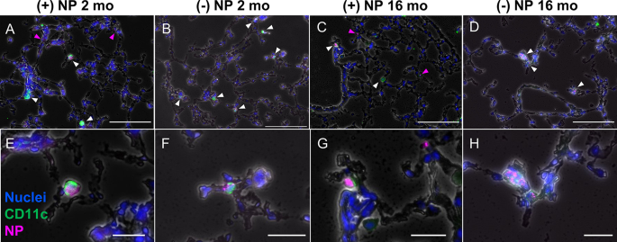
Additional fluorescence histology of frozen lung tissue from consultant mice dosed with NPs for 72 h have been obtained to make sure tendencies of cells aside from AMs have been per stream outcomes. Tissue was handled and imaged to visualise nuclei (DAPI; blue), CD11c (major anti-hamster CD11c; inexperienced), and NPs (pink) Fig. 6). CD11c was used for DC and and to a lesser extent AM isolation. Arrows on particular person photographs level to Cy5-NPs, the place white signifies CD11c+NP+ cells, whereas pink demonstrates NP+ places with out CD11c+ cell current. As anticipated, for each age teams, solely the (+)NP formulation confirmed NPs that weren’t overlapping with CD11c+ cells (Fig. 6A, E, C and G). Since (+)NPs demonstrated a lot fewer AM-specific uptake and all of the interstitial cell sorts exhibited decreased variety of NP+ cells compared to AMs, this means the next probability of “non-associated” NPs within the tissue, which haven’t undergone localization particularly with AMs or DCs. As compared, the (-)NP formulation confirmed larger consistency in uptake of CD11c+ cell throughout each ages with larger prevalence within the 2-month group (Fig. 6B, F, D and H). Not like the BALF photographs nonetheless, there was no constant sample within the variety of NPs that had been phagocytosed between age teams (Fig. 6A and H). That is once more per the truth that DCs uptake NPs in low portions and thus fluorescence imaging might not seize exact adjustments on this regard. These consultant photographs have been per further supplementary information (discuss with Supplemental Determine S10). Thus, these outcomes are additionally consistent with stream cytometry outcomes from Fig. 4.
Immunofluorescence histology highlights CD11c+mobile uptake of NPs. Frozen complete lung murine tissue was sectioned at 10 μm thickness after 72 h dosage of both optimistic (+; A, C,E, G) or adverse (-; B, D,F, H) nanoparticles (NPs; pink) by way of orotracheal instillation. The tissue was stained with major anti-hamster CD11c, secondary IgG Alexa Fluor 488 (inexperienced), and DAPI (nuclei; blue). Consultant photographs (n = 1) characterize 2-month specimens (A, B, E, F) and 16-month specimens (C, D, G, H). White arrows point out overlap between CD11c+, NP+ cells, whereas pink arrows characterize non-overlapping “non-localized” NPs. Pictures acquired utilizing Biotek Cytation 5 Multimode Imager and publicity was modified for viewing of nanoparticles and CD11c stains in every picture. Prime and backside row scale bars characterize 100 μm and 25 μm, respectively
Nanoparticle-mediated phenotype of aged APCs within the lung
As soon as NP uptake was ascertained, the phenotype of the NP+ cells was analyzed to find out if the actual superior age group used right here confirmed indicators of elevated inflammatory potential. H&E evaluation from earlier information of 2-month age group dosed with comparable PEGDA charged NPs had not proven vital upregulation of inflammatory response [38]. Nevertheless, it was notably attention-grabbing to establish if the opposite can be famous within the 16-month group. Consultant H&E photographs confirmed no change in inflammatory expression for both formulation for the aged group, signaling that no main occasion of irritation occurred as a consequence of NP supply (discuss with Supplemental Determine S11). Additional evaluation of the 8 markers used for identification of the 4 APC cell sorts confirmed a rise in CD11b expression for all cells and NP formulations within the aged group after NP uptake, as decided by way of fold change comparability to MFI of younger group for numerous floor markers (discuss with Supplemental Determine S12). Apparently, AMs from the 16-month group confirmed solely a lower in CD11c expression after NP uptake whereas all different cell sorts confirmed a lot higher variation in phenotype following NP uptake (discuss with Supplemental Determine S12). Of observe, all interstitial cell sorts confirmed a rise in MerTK expression and each DC sorts confirmed a rise in CD11c following NP uptake in comparison with the age-matched UT common expression (discuss with Supplemental Determine S12).
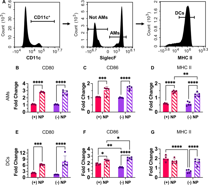
Along with the total stream cytometry panel used to isolate the 4 APCs of curiosity within the lung, an easier panel that used the identical preliminary gating scheme (cells, singlets, dwell cells, CD45+) remoted AMs (CD11c+, SiglecF+) and broadly DCs (CD11c+, SiglecF−, MHC+) (Fig. 7A). This allowed for added comparability of pro-inflammatory floor markers CD80 and CD86 in addition to MHC II after NP uptake. MHC II, a marker of “self” to immune cells that’s vital for antigen presentation to T cells, might develop into activated after NP uptake and maturation of the cell [21, 43, 54]. The untreated specimens introduced totally different CD80, CD86, and MHC II expression between age teams (discuss with Supplemental Determine S13). Outcomes from Fig. 7 characterize age-appropriate fold change information from NP+ cells of every kind permitting for comparability of the rise in inflammatory expression after NP uptake. From this, it was proven that each one inflammatory markers for the 16-month AMs confirmed considerably elevated expression in comparison with the 2-month inhabitants for each the cationic and anionic NP formulations (Fig. 7B and D). Moreover, the (+)NP dosed inhabitants additionally exhibited elevated MHC II expression compared to the (-)NP within the 16-month group AM inhabitants (Fig. 7D). Thus, the upper expression of MHC II for (+)NP formulation right here is consistent with the reducing fee of (-)NP uptake within the aged AM inhabitants.
Nanoparticle uptake in aged cells adjustments activation profile. A.) Multicolor stream cytometry outcomes from partial lung tissue digests after 72-hr pulmonary supply of PEGDA NPs. Alveolar macrophages (AMs; CD45+, CD11c+, SiglecF+) and dendritic cells (DCs; CD45+, CD11c+, MHCIIexcessive) have been remoted from partial lung digest. AMs (B–D) and DCs (E–G) expression of CD80 (B, E), CD86 (C, F), and MHCII (D, G) have been in contrast throughout NP therapies utilizing fold change from age-appropriate UT. Knowledge represents imply ± SD (n = 5). Indicated significance is calculated by way of two-way ANOVA [Tukey Test, p < 0.05 (*), 0.01 (**), 0.001 (***), < 0.001 (****)]
Equally, to the AM inhabitants, the DCs additionally confirmed a rise in CD80 and CD86 expression after NP uptake for every formulation within the aged group (Fig. 7E and F). Of observe, compared to the AMs, the DCs confirmed a lot larger relative fold adjustments for CD80 and CD86 expression for the age inhabitants (Fig. 7E and F). Moreover, each age teams of (+)NP+ samples confirmed a big enhance in CD86 expression compared to the (-)NP+ inhabitants (Fig. 7F). This development was upheld for the 2-month inhabitants for MHCII expression, through which (+)NP formulations have been considerably larger than (-)NP expression; nonetheless, this development was not discovered within the 16-month inhabitants (Fig. 7G). Nevertheless, the elevated inflammatory state within the age mannequin might present a development in direction of over inflammatory responses by these cell sorts; thus correct balancing of protected activation of immunity might be crucial for therapeutics designed for the aged.
